What are Soft Tissue Injuries?
The Periodontist is often called on by the Orthodontist for the exposure of an impacted tooth, which is essential for successful orthodontic treatment. The maxillary and mandibular third molars are the most commonly impacted teeth due to their long development time. The maxillary cuspid is the second most frequently impacted tooth. The cuspids are generally one of the last teeth to erupt into the arch and are adversely affected by:
- The loss of space
- Overretained deciduous teeth
- Deflection (facially or palatally) of the lateral incisor
Various treatment modalities have been proposed to avoid the complications associated with impacted canines. These complications include:
- Internal or external resorption
- Infection associated with partial eruption
- Loss of arch length
- Resorption of the roots of lateral incisors

How are Soft Tissue Injuries caused?
Also, the position of a tooth erupting through the alveolar process in mixed dentition and its ultimate position in relation to the buccolingual dimension of the alveolar process, can have a profound outcome on the amount of attached gingiva around the tooth.
An adequate amount of keratinized gingival tissue that is under proper plaque control, is a fundamental requirement for periodontal health. When teeth erupt uneventfully in the center of the alveolar ridge, an adequate amount of keratinized tissue will surround the erupted permanent tooth. Labially or buccally erupting teeth show reduced dimensions of gingiva as abnormal eruption of permanent teeth restricts or eliminates the keratinized tissue between the erupting cusp and the deciduous tooth. A lack of attached gingiva poses a potential risk for gingival recession in labially or buccally erupted teeth due to the possibility of accumulation of plaque and/or traumatic tooth-brushing during subsequent orthodontic treatment. A good understanding between the orthodontist and the periodontist along with proper management of periodontal tissues can prevent these problems.
Historically, a number of methods have been described for exposure of impacted teeth:
- Celluloid crown
- Pack the wound area to maintain exposure
- Recommended gutta-percha packing
- Pins
- Orthodontic bands
- Wire ligature
The three most significant advances historically for exposure are:
- Palatal flap for exposure
- Direct-bonding brackets
- Soft tissue management
The palatal flap provided access and visibility. Direct bonding reduced morbidity by minimizing wound size and reduced tissue overgrowth and additional surgeries by having a bracket placed at the time of exposure.
Soft tissue management maintained and permitted an increase in keratinized gingiva, eliminating needless secondary surgery to treat mucogingival problems and prevent recession.
Localization and determination of a tooth’s exact position is the foremost step in surgical exposure of an impacted tooth. This can often be done by palpation in labial impactions. However, the use of periapical radiographs and occlusal radiographs plays a major role in palatal and middle alveolar impactions. Use of the buccal object rule is helpful in determining the location of impacted teeth.
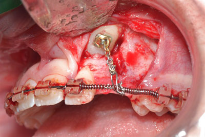
Various surgical techniques can be employed to uncover impacted teeth. The vertical location of the permanent tooth position to deciduous tooth and the amount of gingiva available, will determine the selection of the appropriate technique. The goal of these mucogingival interceptive surgeries is to prevent the ectopic permanent tooth from developing periodontal lesions in its most incipient stage. In this paper, a case report is presented to discuss the validity of utilizing periodontal surgery to increase a band of keratinized tissue in a case of an impacted canine erupting from the alveolar mucosa.
